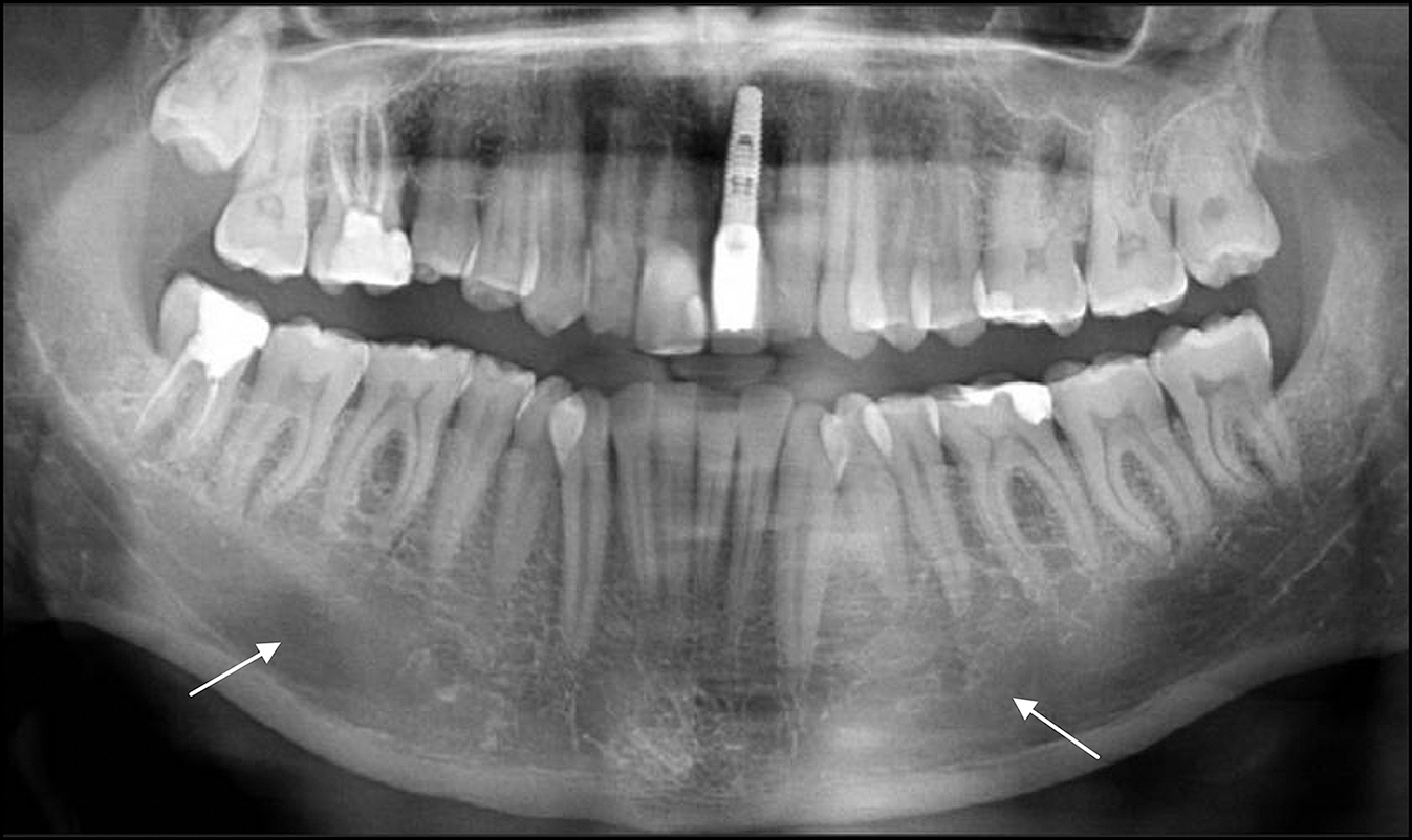
Figure 5.
The orthopantomograph shows the disrupted (arrows) superior border of mandibular canal and cancellous bone which has few and thin trabecullae.
.jpg "View Figure")
.jpg "View Figure")
.jpg "View Figure")
.jpg "View Figure")
.jpg)
.jpg "View Figure")
.jpg "View Figure")