Figure 5. | Photomicrographs showing tumour islands with a peripheral layer of ameloblassts and extensive central areas of granular cells with large, eosinophilic cytoplasmic granules. |
.jpg "View Figure")
.jpg "View Figure")
.jpg "View Figure")
.jpg "View Figure")
.jpg)
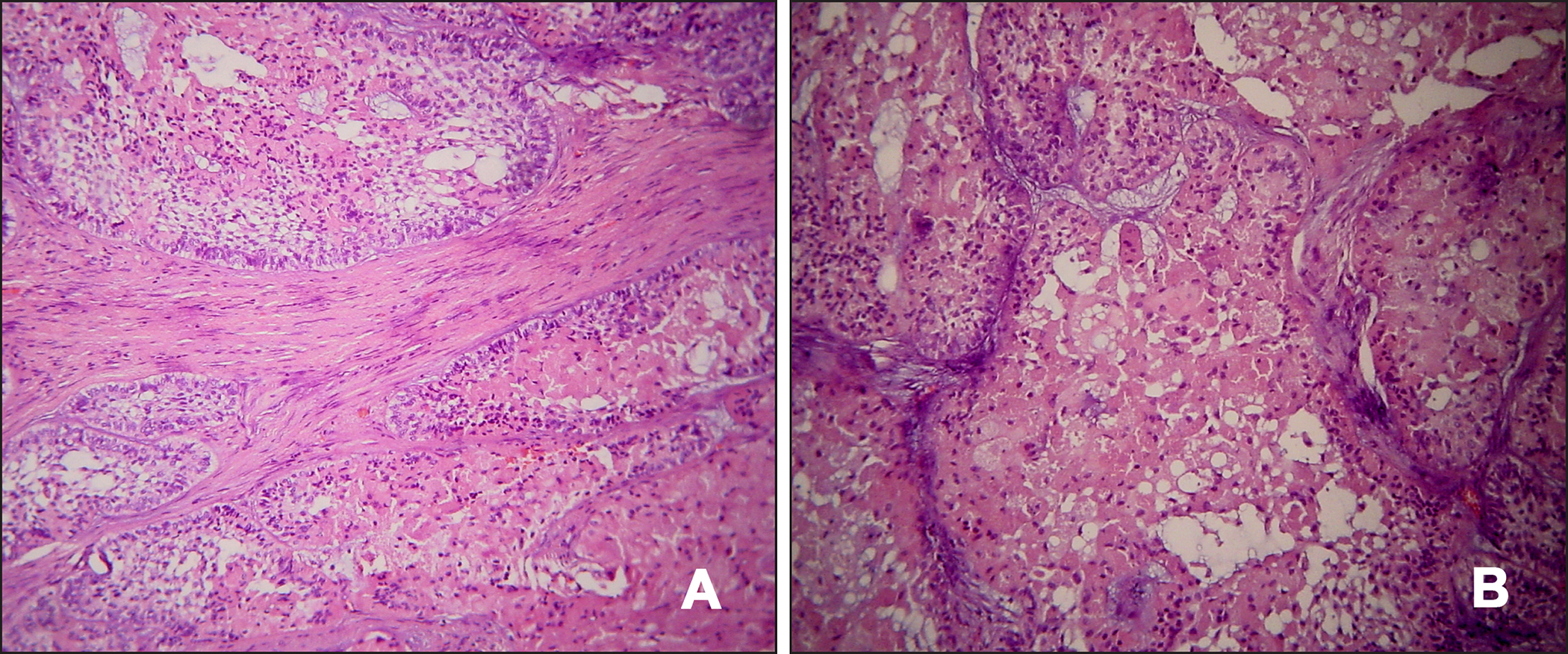
A = hematoxylin and eosin stain, original magnification x100.
|
Figure 5. | Photomicrographs showing tumour islands with a peripheral layer of ameloblassts and extensive central areas of granular cells with large, eosinophilic cytoplasmic granules. |