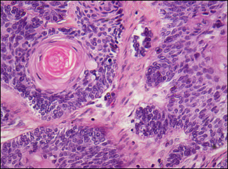
Figure 4.
Haematoxylin and eosin stained section of ameloblastic carcinoma showing cellular atypia (original magnification x400).
.jpg "View Figure")
.jpg "View Figure")
.jpg "View Figure")
.jpg)
.jpg "View Figure")
.jpg "View Figure")
.jpg "View Figure")