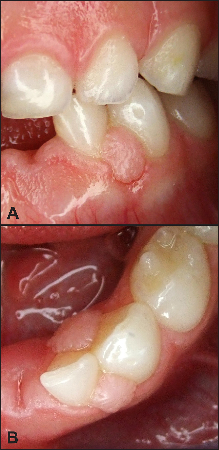
Figure 3.
A = buccal view and B = occlusal view of the nodular lesion of Case report 2 in the interproximal area between the deciduous lateral incisor and canine.
.jpg "View Figure")
.jpg "View Figure")
.jpg)
.jpg "View Figure")