Figure 1. |
A = CBCT image in a sagittal view showing the presence of periapical radiolucency on the apical aspect of the mesial root of a mandibular first molar.
|
.jpg)
Figure 1.jpg "View Figure")
Figure 2
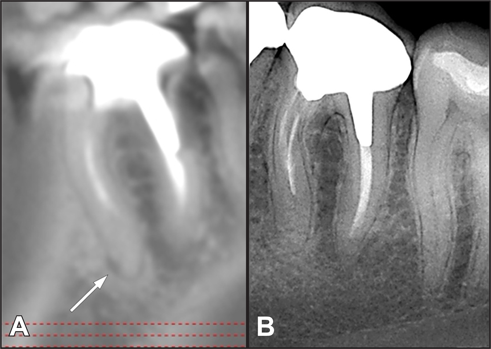
Figure 1. |
A = CBCT image in a sagittal view showing the presence of periapical radiolucency on the apical aspect of the mesial root of a mandibular first molar.
|