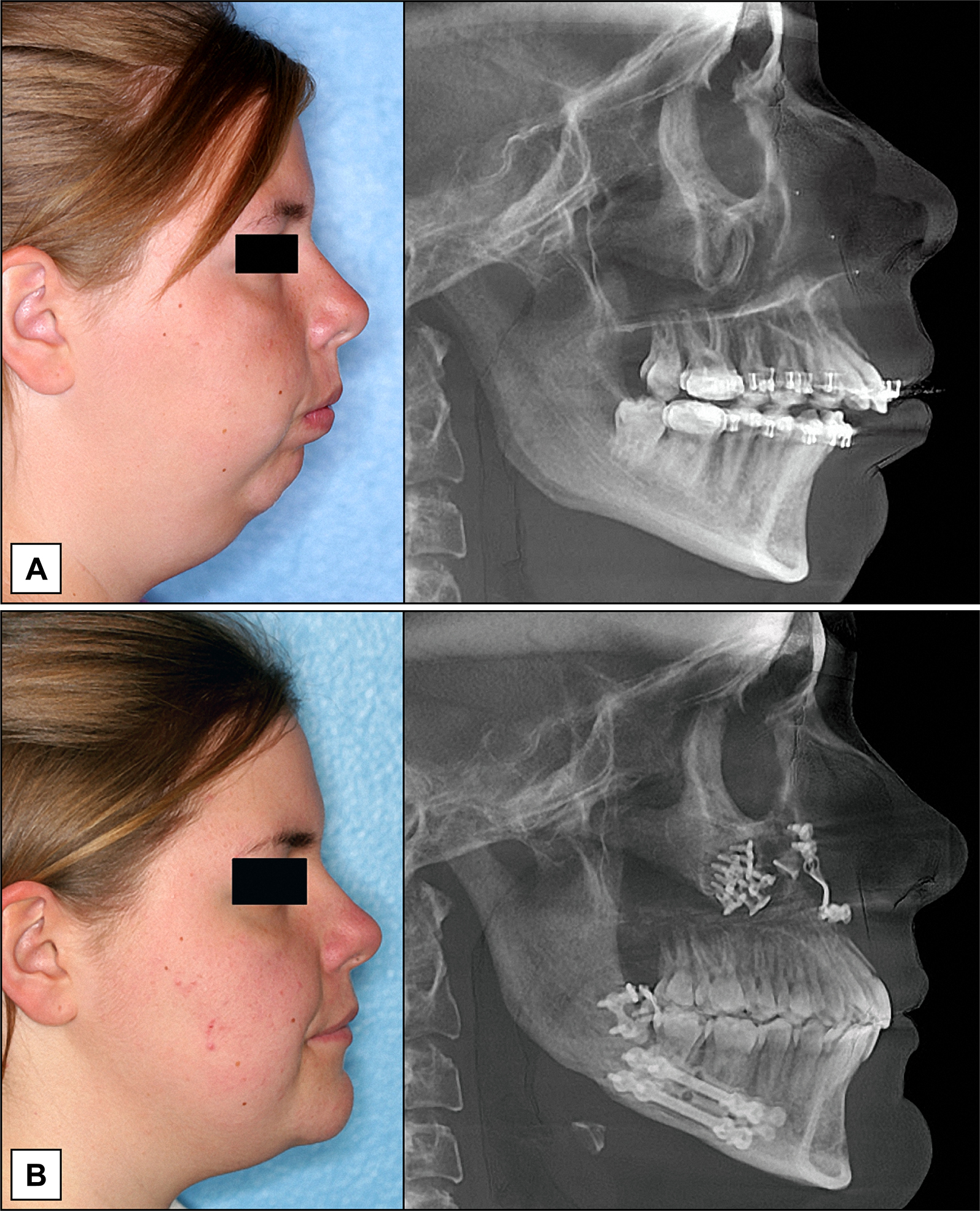
Figure 3.
Clinical and radiographic pictures of a patient treated with bimaxillary orthognathic surgery for correction of a skeletal Class II malocclusion. A = preoperative; B = 18 months postsurgery.
.jpg "View Figure")
.jpg "View Figure")
.jpg)
.jpg "View Figure")
.jpg "View Figure")