Figure 2. |
Dense, diffuse infiltration of a minor salivary gland by a monotonous population of lymphoid cells, destroying the normal architecture.
|
.jpg "View Figure")
.jpg)
.jpg "View Figure")
.jpg "View Figure")
.jpg "View Figure")
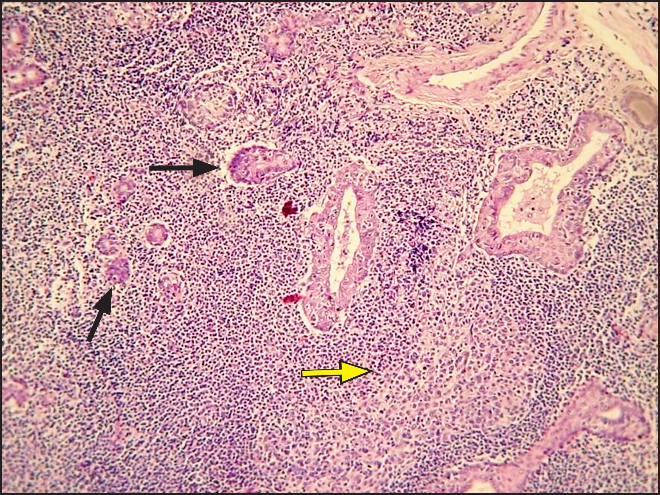
Figure 2. |
Dense, diffuse infiltration of a minor salivary gland by a monotonous population of lymphoid cells, destroying the normal architecture.
|