Figure 3. |
A = histopathologic examination revealed epithelial atrophy, loss of rete pegs, hyalinization of the lamina propria and deep lymphohistiocytic inflammation (hematoxylin and eosin stain, original magnification x100).
|
.jpg "View Figure")
.jpg "View Figure")
.jpg)
.jpg "View Figure")
.jpg "View Figure")
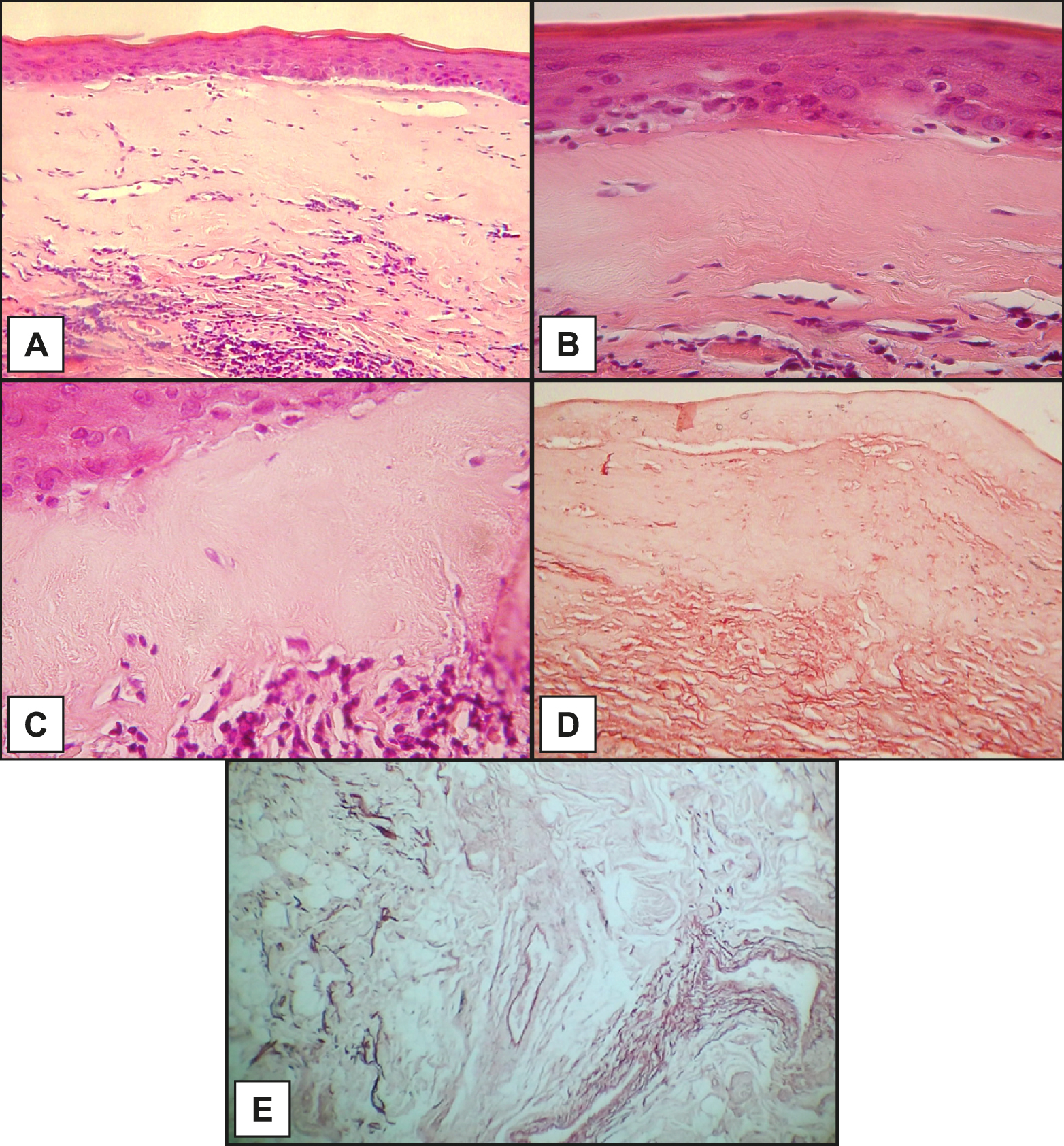
Figure 3. |
A = histopathologic examination revealed epithelial atrophy, loss of rete pegs, hyalinization of the lamina propria and deep lymphohistiocytic inflammation (hematoxylin and eosin stain, original magnification x100).
|