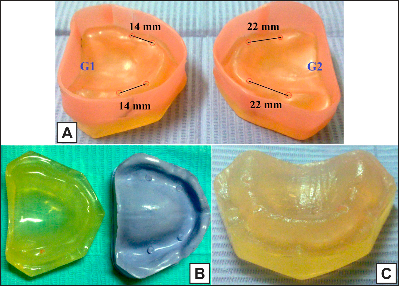
A = maxillary acrylic models waxed and boxed with 3 mm wax free circles at predetermined implant sites.
B = transparent acrylic model (left) and stone index with four stoppers (right) after separation and wax elimination.
C = the acrylic model covered with even thickness silicone layer representing oral mucosa.
.jpg)
.jpg "View Figure")
.jpg "View Figure")
.jpg "View Figure")