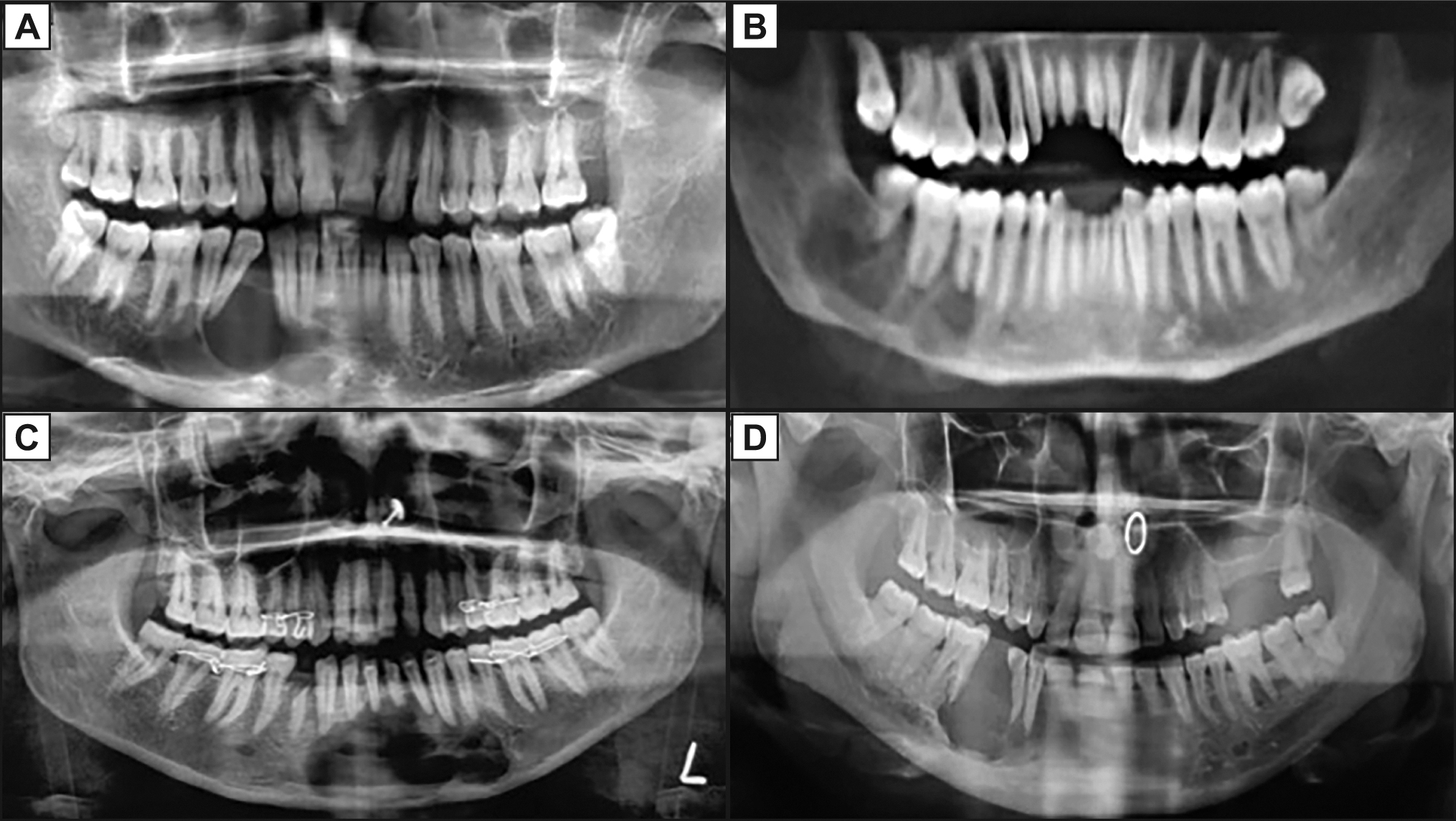
Orpthopantographs depicting:
A = unilocular radiolucency with well defined margins and displacement of tooth;
B = multilocular radiolucency;
C = mixed radiopaque and radiolucent lesion crossing the midline with root resorption and displacement of tooth;
D = radiolucency with displacement of tooth.
.jpg)
.jpg "View Figure")