Figure 1. | Airway analysis was conducted on cone-beam computed tomography scans with Invivo™ software. |
.jpg)
.jpg "View Figure")
.jpg "View Figure")
.jpg "View Figure")
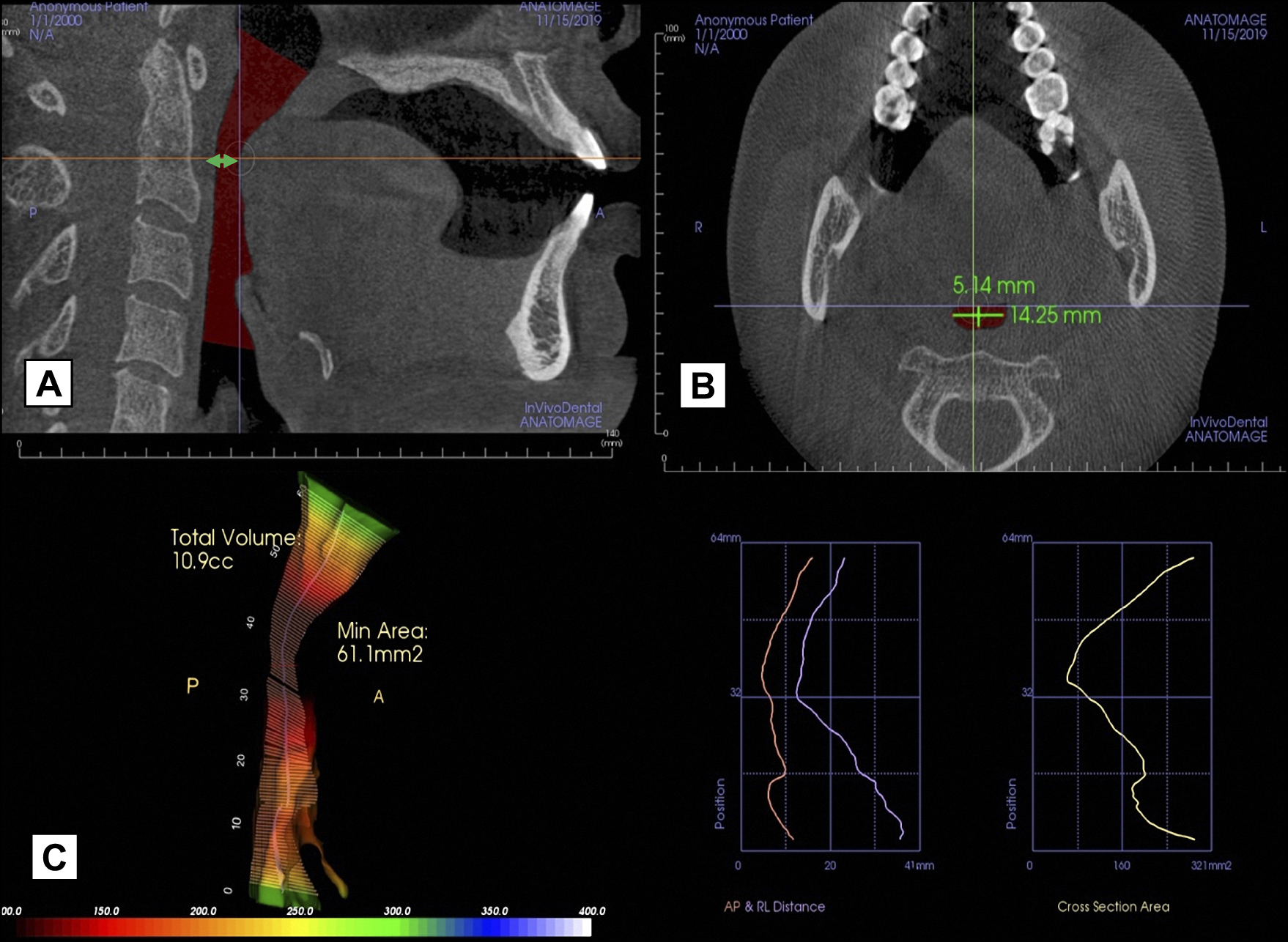
A = on sagittal view, the most constricted point of oropharynx was identified manually, as marked by the green double-headed arrow.
|
Figure 1. | Airway analysis was conducted on cone-beam computed tomography scans with Invivo™ software. |
A = on sagittal view, the most constricted point of oropharynx was identified manually, as marked by the green double-headed arrow.
|