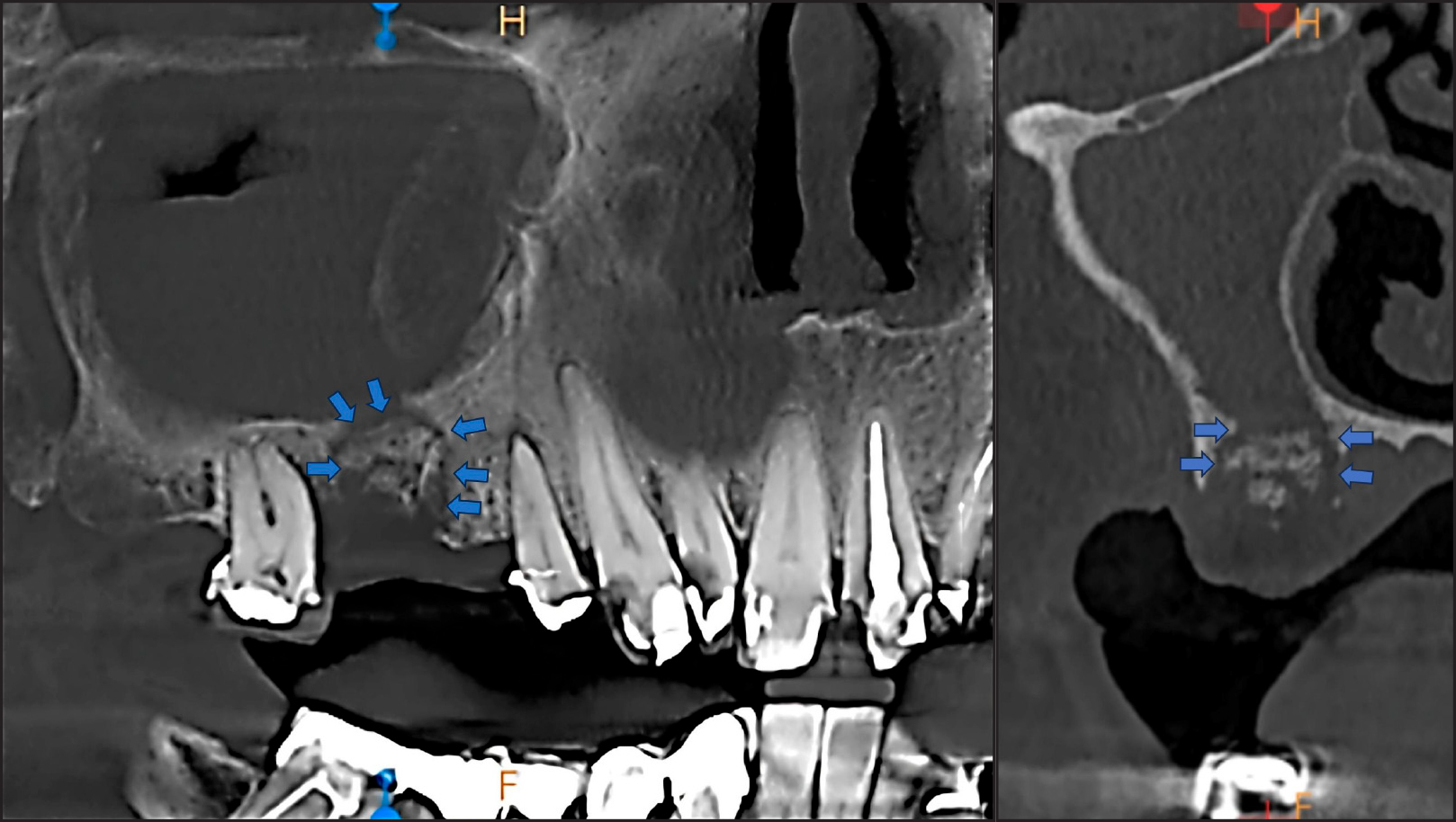
Figure 2.
Initial cone-beam computed tomography examination. Blue arrows indicate the bone lesion. Almost complete opacification of the sinus is visible.
.jpg "View Figure")
.jpg)
.jpg "View Figure")
.jpg "View Figure")