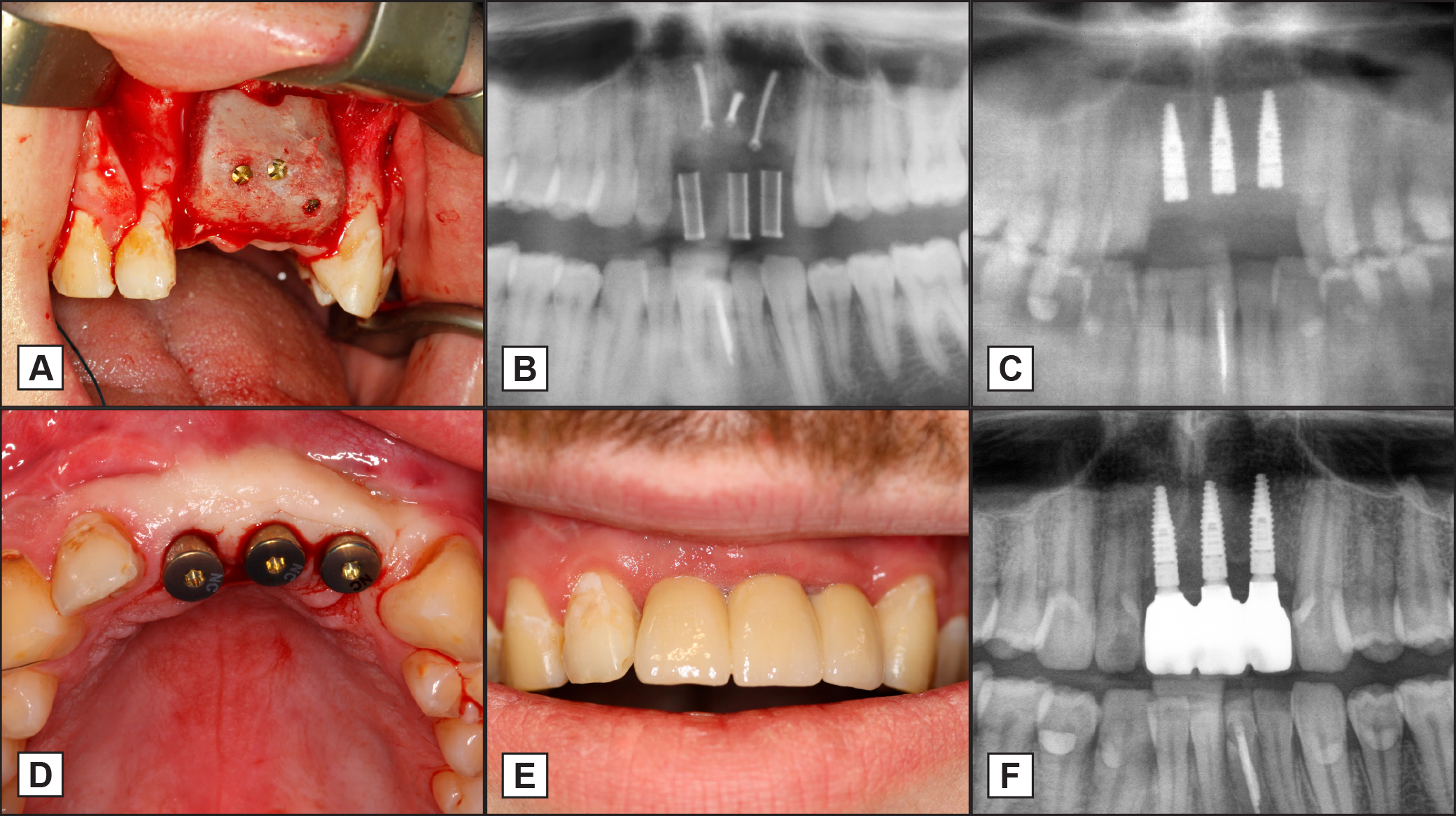
A = maxillary bone augmentation was performed in the anterior area.
B = after a three-month healing period, implant placement was planned and a radiographic control was conducted. C = all three implants were inserted at areas 11, 21, and 22.
D = after a five-month integration time, the implant exposure was performed, and healing caps were added. E and F = clinical and X-ray pictures after three years.
.jpg "View Figure")
.jpg)
.jpg "View Figure")