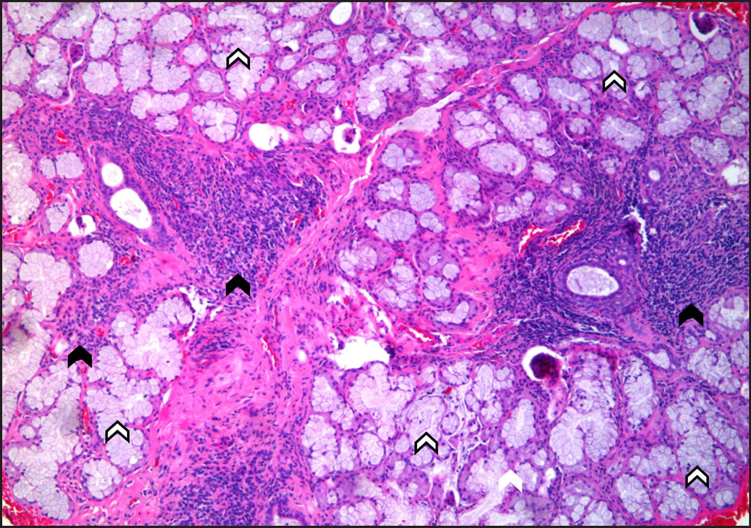
Figure 2.
Labial salivary gland biopsy consistent with Sjögren's syndrome, showing multiple lymphocytic foci (black arrow) and intact acinar units (white arrow) (hematoxylin and eosin stain, original magnification x100).
.jpg "View Figure")
.jpg)
.jpg "View Figure")
.jpg "View Figure")
.jpg "View Figure")