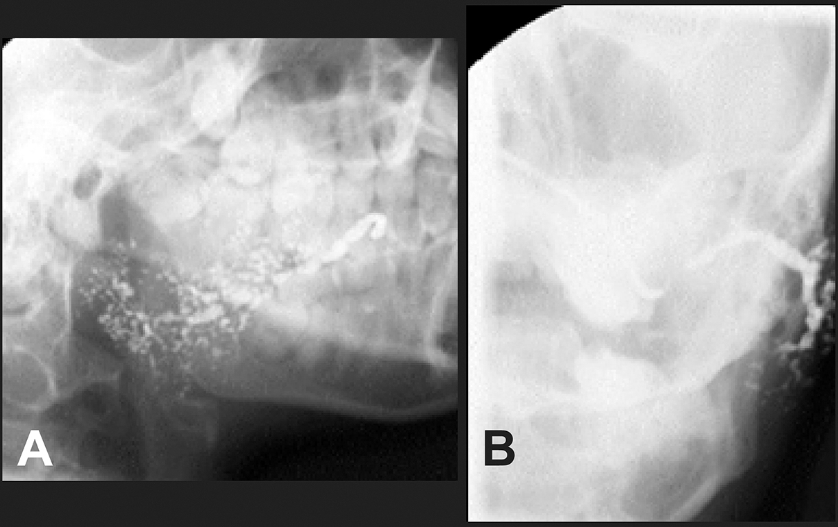
Figure 3.
Sialographic imaging of the parotid gland (Stage 3, according the Rubin and Holt classification).
A = frontal view; B = lateral view. Note the dilated main duct and the sparse overall branching pattern of the ducts.
.jpg "View Figure")
.jpg "View Figure")
.jpg)
.jpg "View Figure")
.jpg "View Figure")