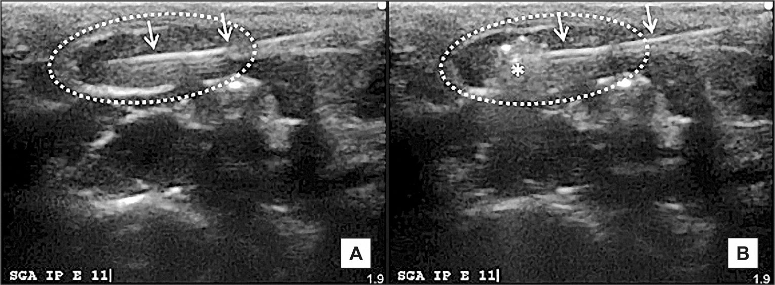
Figure 1.
Ultrasound images demonstrate (A) the advancement of the needle, indicated by double white arrows, and (B) the deposition of 98% ethanol, marked with (*), within the parenchyma of the right SMG, outlined with a dotted line.
.jpg)
.jpg "View Figure")
.jpg "View Figure")
.jpg "View Figure")
.jpg "View Figure")