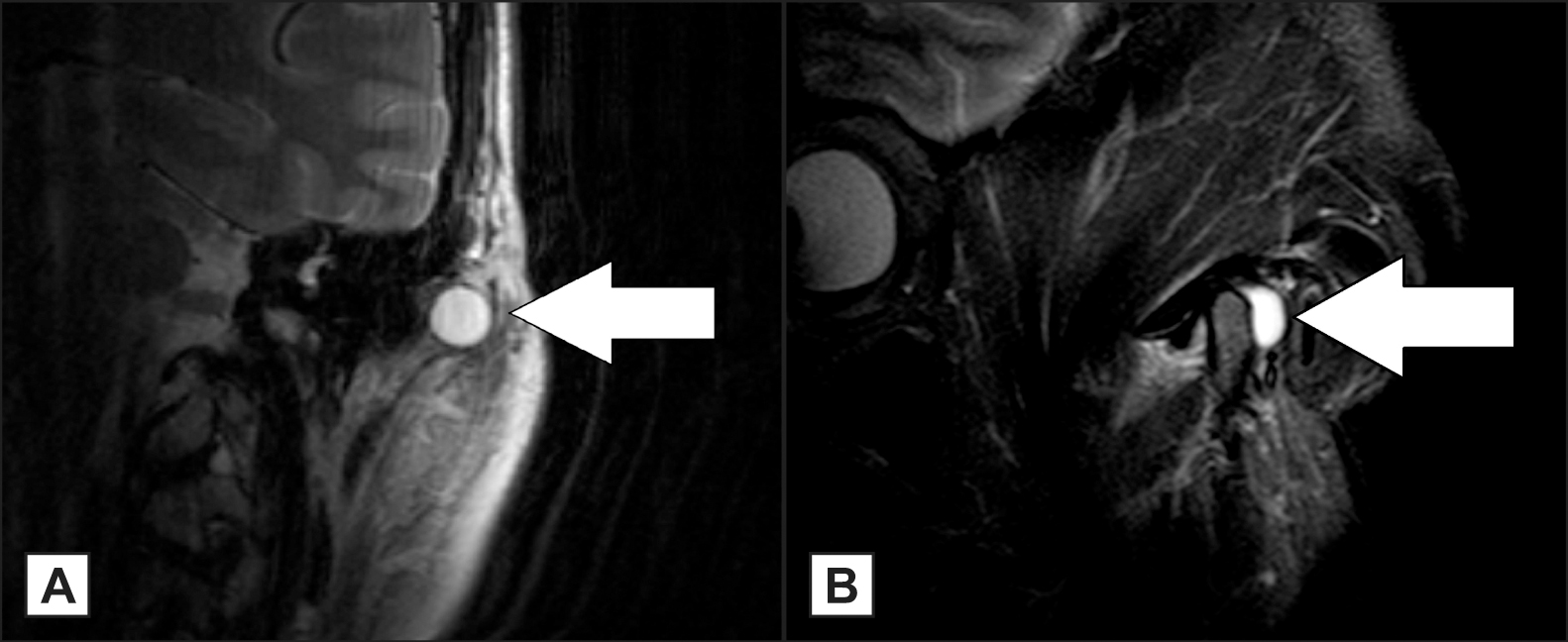
Figure 1.
Preoperative T2-weighted magnetic resonance image. Magnetic resonance imaging of the synovial cyst in the left temporomandibular joint. A = the synovial cyst is visualized in the coronal plane. B = the synovial cyst is visualized in the axial plane.
The cyst is marked by a white arrow. The cyst lumen is fluid filled, making the cyst lumen bright on the T2-weighted MRI.
.jpg)
.jpg "View Figure")
.jpg "View Figure")
.jpg "View Figure")