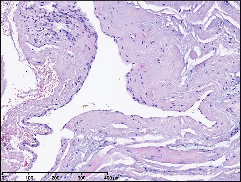
Figure 4.
Histology shows a folded cystic structure lined by flattened synovial cells (original magnification x100).
In the cyst wall myxoid degeneration is observed (hematoxylin and eosin staining).
.jpg "View Figure")
.jpg "View Figure")
.jpg "View Figure")
.jpg)